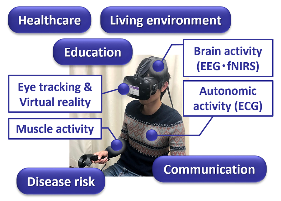
Details of the Initiative
When you become nervous, even though you tried to keep your face calm, a physical response occurs such as your heart beating fast and your hands sweating. Detecting the heart rate and electrical resistance of the skin with a wearable device allows the tension level of the person to be quantified, similar to measuring body temperature and/or blood pressure. Our laboratory is studying to evaluate the invisible intention and emotion from measurements of brain and biological activity.
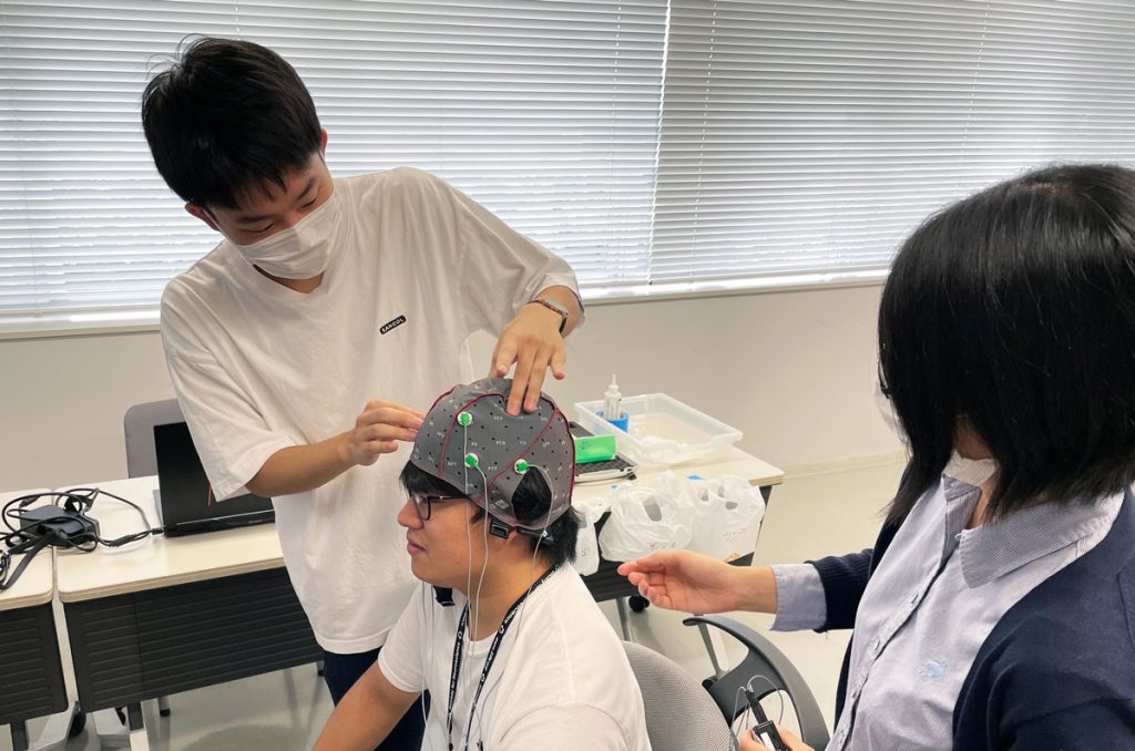
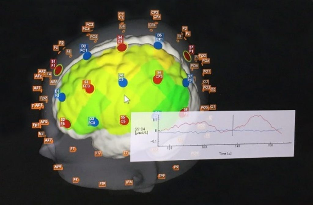
This approach of evaluating anxiety when an earthquake occurs is a collaboration with the Health Science & Medical Engineering Laboratory of the Department of Electronics and Bioinformatics specializing in biometric engineering, the Structural Mechanics & Engineering Laboratory (Professor Masahito Kobayashi), and the Architecture & Structural Engineering Laboratory (Senior Assistant Professor, Tetsuya Tomizawa) of the Department of Architecture. In addition to the conventional physical evaluation of how a building structure shakes in response to earthquake motion, such as acceleration and displacement, we are also using an EEG (electroencephalography) to clarify how much anxiety a person perceived. The establishment of a technology that evaluates anxiety in living spaces will not only contribute to the development of a more user-friendly building structure, but is also expected to be applied to the creation of comfortable spaces and to an indicator of healthcare. Our project has the potential to bring about innovative changes in our lives.